Cardiac Arrest
Objectives
Upon finishing this module, the student will be able to:
- List the two most common causes of pediatric cardiac arrest.
- Identify sick pediatric patients using the pediatric assessment tool (PAT), Primary, and Secondary Surveys.
- Describe pediatric CPR techniques and correct chest compression to ventilation ratios.
- Describe medications used for the treatment of cardiac arrest.
- Describe how to use the Broselow Tape.
- Describe various rhythms seen in cardiac arrest.
- Describe how to monitor a patient after a cardiac arrest.
Contributors
Update Author: Danielle A. Sultan, DO.
Original Authors: Shannon MacRitchie, DO; and Daniel Mielnicki, MD.
Update Editor: Jeff Druck, MD.
Original Editor: S. Margaret Paik, MD.
Last Updated: August 2024
Introduction
Significant advancements in pediatric resuscitation training started in the 1980s. The American Heart Association offers courses in pediatric advanced life support (PALS), which prepares providers with the knowledge and skills to manage critically ill infants and children effectively.
Unlike adult cardiac arrest, which is primarily caused by coronary artery disease, the most common causes of pediatric cardiac arrest are respiratory failure and shock. Timely recognition and management of these disease processes are essential goals of pediatric resuscitation.
EMS brings in a two-year-old male for difficulty breathing and brief loss of consciousness. EMS reports that the child was found in his room this morning by his mother and father with difficulty breathing and then went unconscious for about 20 seconds. He regained consciousness on his own but continued to have difficulty breathing.
EMS arrived and found him hypoxic on room air to 80%, which improved with 10L of oxygen through a non-rebreather mask. Mom and Dad arrive with EMS and tell you that their child had been battling what they thought was a simple cold for the past three days. He had been feeling ill with fevers, runny nose, and a cough. He has not been eating or drinking as much over the past few days.
Start by obtaining a quick general global assessment using the pediatric assessment triangle (PAT). This tool helps to quickly evaluate the status of the child's appearance, work of breathing, and circulation to the skin. Often, you can glean this information within seconds of entering the room.
Appearance
- What is the child's tone?
- How interactive are they with the examiner?
- If they are crying, are they easily consoled?
- Are they looking about the room normally?
- Is their cry or speech normal?
Work of Breathing
- Does breathing look easy?
- Are there signs of respiratory distress such as accessory muscle usage, subcostal and intercostal retractions, tracheal tugging, head bobbing, or nasal flaring?
- Is the child tripoding?
- Is the child gasping or apneic?
Circulation to Skin
- Is the color of the skin normal?
- Is there presence of pallor, cyanosis, or mottling?
- What is the skin temperature (i.e. warm or cold)?
The assessment should take no more than a few seconds. Any abnormalities in these areas should give concern about the patient's stability and prompt swift action.
Primary Survey
The primary survey consists of basic assessments of systems that significantly impact sustaining life. An abnormality in one of these areas is considered a threat to life and should prompt immediate intervention. Ensure your primary survey includes vital signs to identify instability.
Airway: Is the Airway Patent?
In children, you can assess this by seeing if the child is able to speak. In younger children who have not learned to speak, you can see if they are crying, babbling, or making sounds from the mouth. Always make sure to look inside the mouth for obstruction or foreign body.
Breathing: Is the Child Breathing?
Take another moment to notice signs of respiratory distress as listed above. You should auscultate the chest, make sure there are lung sounds on both sides of the chest, and listen for abnormal breath sounds.
Circulation: Does the Child Have Signs of Good Perfusion?
Pediatric patients can compensate really well for low blood pressure. A normal blood pressure reading does not always indicate that they have good perfusion. Do they have a strong or weak pulse? Is the skin on their extremities warm or cool? Is their skin dry or clammy? Is there good skin turgor?
Disability: What is the Child's Level of Consciousness?
You can use AVPU to determine the level of consciousness in a pediatric patient.
- Alert - check to see if the child is alert.
- Voice - does the child respond to verbal stimuli?
- Pain - does the child respond to painful stimuli?
- Unresponsive - is the child unresponsive
The secondary survey can tell you a lot about the root cause of a patient's condition. Perform a rapid but thorough exam from head to toe and look for abnormalities. Afterward, you can try to get a brief history from either the patient or their parent/guardian/caretaker using the SAMPLE mnemonic: signs and symptoms of presenting complaint, allergies, medications, past medical history, last meal, and events leading to current illness.
CASE STUDY: Secondary survey reveals sunken eyes, clear rhinorrhea, and dry/cracked lips. His pharynx is erythematous but there are no tonsillar exudates or enlargement. The patient is coughing and spits up yellow/tan mucous. There are wheezes throughout his lungs and coarse rhonchi in the right lower lobe. His abdomen is soft and nontender, with no abnormalities in the GU area. He does not have a rash. Initially he had been moving all of his extremities, but now appears to lay limp on the stretcher.
- Subjective: Mom tells you he has been sick for three days with a runny nose, cough, and fever as high as 102.5F. He had decreased appetite over the past day and she can't remember the last time she changed a diaper.
- Allergies: none.
- Meds: none.
- Past Medical History: full term, no birth complications, no stay in the neonatal ICU.
- Last Meal: could only drink three ounces of milk last night.
- Events: found him this morning with difficulty breathing.
As you are performing your assessment and gathering history, your patient's eyes roll back and the nurse notes he stops breathing. You cannot feel a pulse...
Recognition of cardiac arrest is the first step in Pediatric Advanced Life Support (PALS).
Feeling for a Pulse
Palpation of a pulse occurs in different locations based on the child's age. In school-aged children, start by feeling for a radial pulse. In younger children such as infants and neonates, look for a pulse in the brachial artery. If you cannot feel a pulse in these locations, try to feel one in the femoral artery in both age groups. Searching for a pulse should take no more than ten seconds. Trust your gut - if you don't feel it, then it's not there.
Chest Compressions
If you do not feel a pulse, start chest compressions immediately. For one-rescuer CPR, alternate between 30 chest compressions and two breaths (30:2). For two-rescuer CPR, alternate between 15 chest compressions and two breaths (15:2). The preferred method for chest compressions in infants is to use both hands, encircling the chest with both thumbs and compressing the lower sternum. The alternative technique is using two fingers from one hand to provide compressions on the lower sternum.

Figure: Pediatric CPR techniques.
Effective chest compressions are critical:
- Compression rate should be at least 100 per minute.
- Compression depth should be 1/3 of the anteroposterior diameter of the chest, or 4cm in infants and 5cm in pediatric patients.
- Make sure to allow for complete chest recoil in between compressions.
- Swap out the person doing compressions every two minutes to avoid fatigue.
Note: while chest-compression-only CPR is an effective initial treatment for cardiac arrest in adults, compression-only CPR is not recommended for infants and children. This is because cardiac arrest in children is most commonly caused by hypoxia, and providing rescue breaths is crucial for reversing the most likely cause of arrest.
Oxygenation and Ventilation
When a patient is in cardiac arrest, they are not breathing on their own and it becomes your job to oxygenate and ventilate them appropriately. Initially, this is begun by using a bag-valve-mask (BVM), in which a mask is placed over the patient's mouth and nose, forming a tight seal. Oxygen is delivered into the lungs as the bag is squeezed. Again, the rate of breaths should be 30 compressions to 2 breaths in one-person CPR and 15 compressions to 2 breaths in two-person CPR.
While performing PALS, consider obtaining a definitive airway by intubating the patient and putting in an endotracheal tube. This helps support the patient's oxygenation and ventilation while they are not responsive and cannot breathe on their own. Once a patient has a definitive airway, you no longer have to pause compressions to deliver breaths. Instead, compressions can be administered ceaselessly (aside from stopping to check the patient's rhythm) while breaths are delivered every six seconds.
Medications in Pediatric Cardiac Arrest
In pediatrics, most medications are dosed based on the patient's weight. However, in emergency situations, there is often not enough time to weigh the patient. PALS supports using the Broselow Pediatric Emergency Tape to reduce errors involved in calculating correct dosages. The tape displays doses for common emergent medications based on a child's length, making it easy and quick to use. To use the tape:
- Place the part of the tape marked "Start" at the head of the patient.
- Extend the tape down the length of the patient.
- Evaluate which color corresponds with the heels of the patient.

Figure: Broselow's Tape.
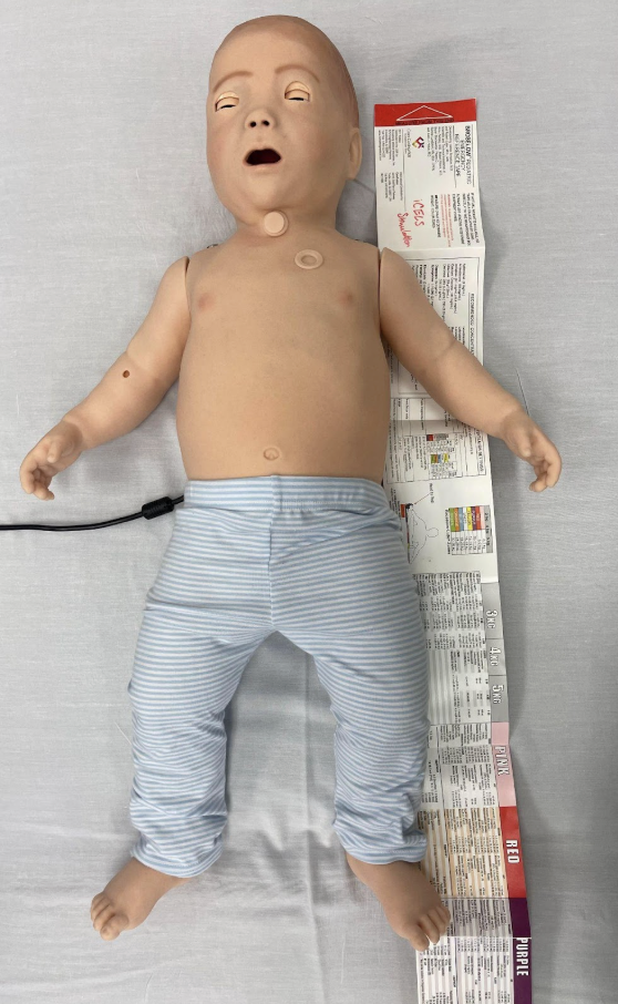
Figure: Measuring child using Broselow's tape.
Use the appropriate color section on the tape to identify the appropriate weight-based dosing of medications, equipment sizes, and other life-saving medications.

Figure: Details shown in correlated color of Broselow's tape.
Note: The Broselow tape is designed to help providers determine approximate medication doses in emergencies such as cardiac arrest. If your patient is more stable and you can get an accurate weight, this is the preferred method of dosing medication.
Epinephrine
Regardless of the initial cause, epinephrine is the treatment for cardiac arrest. The dose of epinephrine is 0.01 mg/kg (or 0.1 mL/kg) of the 1:10,000 concentration repeated every three-five minutes. If the initial dose is ineffective, additional doses should be given at the same dose. Epinephrine is most often given through an IV. If there is difficulty in establishing IV access, an intraosseuous (IO) line can be placed to administer medications. In situations where IV/IO access is unavailable, epinephrine 1:1,000 can be given via the endotracheal tube. The dose is 0.1 mg/kg (or 0.1 mL/kg). A normal saline flush is instilled after the epinephrine. It can also be given through a central line or intramuscularly.
Rhythms in Cardiac Arrest
While performing PALS, you should routinely and often check for the child's response to your interventions. Perform rhythm checks every two minutes to identify rhythms that are potentially shockable or require defibrillation. Below are types of rhythms you may find on the rhythm checks in a cardiac arrest.
- Asystole: Asystole occurs when the heart's electrical activity is not discernible and there is no pulse. The treatment for asystole is to continue high-quality CPR and PALS.
- Pulseless Electrical Activity (PEA): PEA occurs when the heart has organized electrical activity but no pulse. The electrical activity can be a variety of different rhythms, but most often is sinus rhythm. The treatment for PEA is to continue high-quality CPR and PALS.
- Pulseless Ventricular Tachycardia: Pulseless Vtach occurs when electrical activity moves very quickly through the ventricles and often skips the atria. The monitor will have wide, regular QRS complexes. Each QRS may look all the same as in the figure below (monomorphic) or have a variety of different morphologies (polymorphic). The treatment for pulseless vtach is defibrillation. If the rhythm does not improve despite two shocks, consider administering an antiarrhythmic such as amiodarone or lidocaine.
- Ventricular Fibrillation (VFib): VFib occurs when the ventricles are attempting to fire, sometimes up to 500 bpm! This makes organized ventricular contractions impossible, resulting in a drop in cardiac output. On the EKG or monitor, you will see chaotic irregularities without an obvious QRS morphology. The treatment for Vfib is defibrillation. If you have no improvement of the rhythm despite two shocks, consider administering an antiarrhythmic such as amiodarone or lidocaine.

Figure: Monomorphic ventricular tachycardia. Image courtesy of E. Burns and R. Butler. Used under the Creative Commons Attribution-NonCommercial-ShareAlike 4.0 International License.

Figure: Polymorphic ventricular tachycardia. Image courtesy of E. Burns and R. Butler. Used under the Creative Commons-Attribution-NonCommercial-ShareAlike 4.0 International License.

Figure: Ventricular fibrillation. Image courtesy of E. Burns and R. Butler. Used under the Creative Commons-Attribution-NonCommercial-ShareAlike 4.0 International License.
How to Defibrillate
- Make sure you place both pads on the child's chest. The recommended placement of pads in pediatric patients is anterior-posterior, where the anterior pad is placed over the sternum and the posterior pad is placed over the mid-thoracic spine.
- Turn on the defibrillator and switch to manual mode.
- Choose the amount of energy you want to deliver. Your first defibrillation should be approximately 2 Joules/kg, the second defibrillation should be approximately 4 Joules/kg, and every defibrillation thereafter should be 4 Joules/kg.
- Press the charge button and continue chest compressions while the defibrillator is charging. The machine will alarm once it is fully charged.
- Make sure everyone on your team moves away from the patient and does not touch them. Most people shout, "clear!"
- Press the shock button to deliver the shock.
- After each shock, you must resume chest compressions and PALS until the next rhythm check.
Labs and Ancillary
It is important to determine the potential cause for the arrest as a means of treating the underlying cause of arrest as well as preventing subsequent arrests. Cardiac arrest causes are typically thought of using the H's and T's:
- H: Hypoxia, hypoglycemia, hypo/hyperkalemia, hypothermia, hypovolemia, H+ (acidosis).
- T: Trauma, thrombosis (pulmonary embolism), thrombosis (myocardial infarction), tension pneumothorax, tamponade, toxins.
Your workup of a patient who has had cardiac arrest should largely be focused on identifying the underlying cause of their arrest, as well as complications from performing advanced life support. You can order tests and perform studies to evaluate the presence of or exclude certain diagnoses in patients. For example, below is a list of tests that can be ordered and how they can evaluate a diagnosis:
- Complete Blood Count: White blood cells (can indicate severe infection), hemoglobin/hematocrit (can indicate excessive blood loss).
- Comprehensive Metabolic Profile: Abnormal electrolytes can indicate hypo/hyperkalemia.
- Blood Gas (arterial or venous): Can evaluate for acidosis or significant hypoxia.
- Chest X-ray: Can look for pneumothorax, source of hypoxia such as pneumonia or acute respiratory distress syndrome, heart failure, or congenital heart disease.
- Point-of-Care Ultrasound (heart, lungs, focused trauma assessment): Heart failure or heart attack, cardiac tamponade, massive pulmonary embolism, pneumothorax or pulmonary edema, hypovolemia from intra-abdominal free fluid in the setting of trauma.
- Toxicology Panel: Identify toxicologic substances that may have led to hypoxia and arrest or dysrhythmia during arrest.
- Point-of-Care Glucose Test: Identifies hypoglycemia.
- EKG: Can identify abnormalities in conduction that can lead to arrest such as Wolffe Parkinson White, congenital long QT syndrome, or complete heart block.
- Troponin: Can indicate myocardial infarction.
- D-dimer: Can be used to risk stratify the presence of pulmonary embolism.
The most common reason for a pediatric patient to suffer a cardiac arrest is from hypoxia or respiratory arrest.
CASE STUDY: You start two-person CPR on the patient, where the compressor has their hands in a two-thumb encircling technique. Breaths are delivered at a rate of two breaths every 15 compressions via bag-valve mask. You rapidly intubate your patient with the appropriately-sized equipment and confirm placement of your endotracheal tube by listening to the lungs and using a color-change colorimeter. You instruct your team that now they can deliver breaths every six seconds and continue CPR without stopping. The patient falls into the red category on the Broselow's Tape, so you search the listed medications and find the approximate weight-based dosing for life support medications, administering 0.085 mg (or 0.85 mL) of cardiac epi. You analyze the child's rhythm and find a thin wavy line that is variable and does not have any discernible QRS complexes. However, it is also not a flat line. You identify this as ventricular fibrillation and appropriately administer defibrillation. After you administer defibrillation and continue CPR for another three minutes, it is time for another rhythm and pulse check. You find that the child has a perfusing rhythm of sinus tachycardia and your nurse identifies a pulse in the femoral artery. You have achieved ROSC.
With the help of high-quality PALS, you will hopefully obtain return of spontaneous circulation (ROSC, pronounced “Raw-sk”). Although you’ve done an excellent job saving your patient’s life, the patient still needs your help to continue to provide advanced life support and address immediate life threatening conditions.
In the initial phase, repeat vital signs and re-perform the primary and secondary surveys. Cardiac arrest and CPR can cause serious adverse consequences including rib fractures, pneumothorax, intra abdominal organ lacerations, myocardial injury, tracheal injury, and brain injury from hypoxia. Maintain adequate blood pressure and perfusion with IV fluid boluses and/or vasopressors as needed. Reevaluate potential causes of the initial cardiac arrest and treat them immediately if they are reversible.
CASE STUDY CONCLUSION: You admit the patient to the pediatric intensive care unit. It was determined they had a significant viral illness causing bronchiolitis and subsequent respiratory failure and cardiac arrest. They were extubated on day three of their hospital admission and went on to have a full recovery without any neurologic or cardiac damage.
- Cardiac arrest in the pediatric population is most often secondary to respiratory arrest and shock. Recognizing ill children early and intervening quickly improves outcomes.
- Pediatric patients should receive both chest compressions and ventilatory support rather than chest compression-only CPR.
- Weight-based resuscitation tapes can be a valuable resource for critically ill infants and children, and the Broselow tape can help rapidly identify doses of emergency medications.
- It is essential to assess and treat the underlying cause of the cardiac arrest.
- Web-Based Integrated Guidelines for Cardiopulmonary and Emergency Cardiovascular Care - Part 12. Pediatric Advanced Life Support. American Heart Association.
- Burns E, Butner R. Ventricular Fibrillation. Life in the Fast Lane. 2023 Mar 24.
- Burns E, Butner R. Monomorphic Ventricular Tachycardia. Life in the Fast Lane. 2023 Mar 19.
- Burns E, Butner R. Polymorphic Ventricular Tachycardia. Life in the Fast Lane. 2023 Apr 15.
- de Caen AR, Berg MD, et al. Pediatric Advanced Life Support: 2015 Guidelines Update for Cardiopulmonary Resuscitation. American Heart Association.
- Lopez-Herce J, Garcia C, et al. Outcome of Out-of-Hospital Cardiorespiratory Arrest in Children. Pediatr Emerg Care. 2005.
- Emergency Cardiovascular Care. Circulation. 2015.
- Schindler MB, Bohn D, et al. Outcome of Out-of-Hospital Cardiac or Respiratory Arrest in Children. N Engl J Med. 1996.
- Young KD, Seidel JS. Pediatric Cardiopulmonary Resuscitation: A Collective Review. Ann Emerg Med. 1999.