Snake Bites
Author: Jessica Slim, MD, MPH, PGY3 in Emergency Medicine, Denver Health Medical Center
Editor: Rahul Patwari, MD
Objectives
Upon completing this module, you should be able to:
- Recognize the clinical presentation of Viperidae and Elapidae snakebites in the U.S.
- Understand the key treatment principles of Viperidae and Elapidae snakebites.
Introduction
The main types of venous snakes in the United States include the Viperidae (Crotalid, pit viper) and Epalidae (coral snake species). Venomous snakes are found throughout the Unites States except in Maine, Alaska, and Hawaii. The majority of bites occur between May and October as most snakes hibernate in the winter. Most bites involve the extremities, but can occur to the face or tongue when the snake is held close to the body. Children, intoxicated persons, snake handlers and collectors are frequent victims. Adult patients typically present with upper extremity injuries and children typically present with lower extremity injuries. Viperidae snakes account for 99% of venomous snakebites in the United States. The remaining 1% result from coral snakes and exotic species. This is because Elapidae venom apparatus is not as efficient for venom delivery and the species mouth is much smaller.
It is important to note that 25% of crotaline bites do not impart venom; these are known as “dry” bites, defined as bites that do not result in local tissue damage, hematological abnormalities, or regional lymph node pain. The true incidence may be much higher since they may not be seen in the emergency department and thus are not reported to the poison control center.
Etiology
Viperidae Snakes
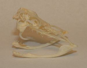
Viperidae snakes, also known as pit vipers, are identified by their heat-sensing pit organs, fangs, triangular head, and elliptical pupils. The fangs are connected to venom sacs that inject venom. The caudal end of the snake has a single row plates or scales, as opposed to the double row of scaled found on non-venomous varieties. Rattlesnakes account for 65% of Viperidae bites; copperheads are responsible for 25%. The remaining 10% are from water moccasins.
Elapidae Snakes
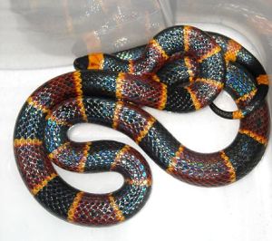
The mnemonic “Red on Yellow, Kill a Fellow. Red on Black, Venom Lack (or Friend of Jack)” applies only to North American coral snakes
The coral snakes have distinctive red, yellow and black bands. There are many mimickers of coral snakes, including the California king snake. The king and coral snake can be distinguished by their colored rings and the color of their snouts. Coral snakes have black snouts and king snakes have red snouts. In the coral snake, the red and yellow rings touch, while in the king snake, the red and black rings touch. This has led to the saying, “Red on yellow, kill a fellow; red on black, venom lack.” Coral snakes are not aggressive and live mostly underground. Thus, most of the bites occur while being handled, often because the victim believes they are dealing with the nonpoisonous California king snake. The snake must chew to deliver its venom. It is thought that the snake must maintain its bite for a prolonged period of time in order to administer a significant amount of venom.
Initial Actions and Primary Survey
In the pre-hospital setting, the affected extremity should be placed in a neutral position and pain control initiated. The initial evaluation in the emergency department is no different for any other patient who presents for evaluation and management. After airway, breathing, and circulation have been evaluated and stabilized, a detailed history should be obtained for the following information: previous comorbidities (e.g. bleeding dyscrasias), medications (e.g. warfarin), immunization status, allergies, type of animal which caused the envenomations, time and location of bite, the progression of symptoms since envenomation, and the types of pre-hospital therapies performed. The affected limb should be elevated (only once the patient has arrived to the emergency department) and the area surrounding the bite should be marked to assess progression of symptoms. Tourniquets should not be applied to extremities with snake bite wounds.
Presentation
Viperidae venom is a complex solution of various proteins, peptides and enzymes that allow the snake to kill its prey quickly and begin the digestive process. These bites usually cause severe pain from the time of envenomation and swelling that can progress at various rates due to lymphatic spread. The bite site may become edematous and tense, develop ecchymosis, fluid filled or hemorrhagic bullae and extensive tissue destruction may eventually develop. Swelling may be marked, but compartment syndrome is rare. The full extent of local or systemic involvement may not be evident for hours. Hematologic abnormalities are common in Viperidae envenomation, including thrombocytopenia, elevated PT, and decreased fibrinogen. Systemic toxicity can result in oral paresthesias, metallic taste, fasciculations, tachycardia, hypotension and anaphylaxis. As a general rule, if there are no symptoms within six to eight hours, the patient can be considered medically cleared.
Elapidae Venom has various toxins which produce systemic neurotoxicity. Coral snake envenomation may present with serious systemic toxicity with little findings at the actual site of envenomation due to the venom’s lack of cytotoxicity. The neurologic abnormalities may include weakness, numbness, fasciculations, tremor, diplopia, bulbar palsies with slurred speech, dysphagia and respiratory paralysis (immediate cause of death). Ptosis is frequently the initial sign of toxicity. Coral snakes have the potential to cause high morbidity with respiratory failure, neurologic dysfunction and cardiovascular collapse, requiring airway and respiratory management lasting several weeks. Onset of clinical effects following envenomation occurs between one and seven hours but may be delayed up to eighteen hours.
Because children have smaller body mass, smaller limbs, and less subcutaneous tissue, they can potentially receive more venom per kilogram body weight and therefore have more clinical severity than adults.
Diagnostic Testing
In cases of Viperidae envenomation, baseline studies including CBC, platelets, PT, PTT, INR, fibrinogen, electrolytes, creatinine and UA should be obtained on ED arrival and repeated in four to eight hours.
Treatment
Aggressive supportive care, including initial assessment of patient’s airway, breathing, and circulation, wound care, and tetanus prophylaxis. Generous use of opioids may be necessary to effectively control the patient’s pain. Antibiotics are usually not necessary. All coral snake bites require admission for observation.
Antivenoms can help correct systemic dysfunction and coagulopathy. They can also help stop progression of further local edema, hemorrhage, and soft tissue swelling if these conditions are treated early. Antivenom is indicated for all eastern coral snake bites (even if asymptomatic). Otherwise, antivenom is indicated based on degree of continued spread or if any systemic symptoms develop. Usually antivenom doses are not adjusted based on a child’s weight or age because the antivenom dosage should reflect venom load rather than patient size. It is important to monitor for recurrence phenomena and rebound coagulopathy following treatment with CroFab antivenom.
Pearls and Pitfalls
Coral snake envenomation presents with delayed neurotoxicity.
- Antivenin may require repeat doses due to recurrence phenomenon.
- Antivenom can cause hypersensitivity reactions and may lead to anaphylaxis.
- Pre-hospital care of viperidae snakebites should be confined to immobilization of the affected limb and neutral position.
- Fasciotomy is rarely indicated for Viperidae snake bites (sub-fascial envenomation is unusual, true compartment syndrome is usually note present, some studies suggest that fasciotomy may worsen outcome).
- Consult your local poison control center early for advice in the course of treatment.
Selected References
- Cheng, AC. Management of Crotalinae (rattlesnake, water moccasin [cottonmouth], or copperhead) bites in the United States. In: UpToDate, Post TW (Ed), UpToDate, Waltham, MA. (Accessed August 1, 2014).
- Cheng, AC. Management of Elapidae (coral snakes) bites in the United States. In: UpToDate, Post TW (Ed), UpToDate, Waltham, MA. (Accessed August 1, 2014).
- Feng, S and Goto, C. Bites and Stings – Snakes, Spiders, and Scorpions in the United States. Emergency Medicine Practice. May 2007. Volume 4, Number 5. (Accessed from ebmedicine.net August 1, 2014).
- Jacobstein, C.R. and Baren, J.M.: Bites and Envenomations (Section XXIV Environmental Emergencies) in Wolfson, A.B., Hendey G., et al. (Eds.). Harwood-Nuss’ Clinical Practice of Emergency Medicine, (5th edition), Lippincott, Williams and Wilkins, Philadelphia, 2009.
- “Coral 009” by Norman.benton – Own work. Licensed under CC BY-SA 3.0 via Wikimedia Commons.